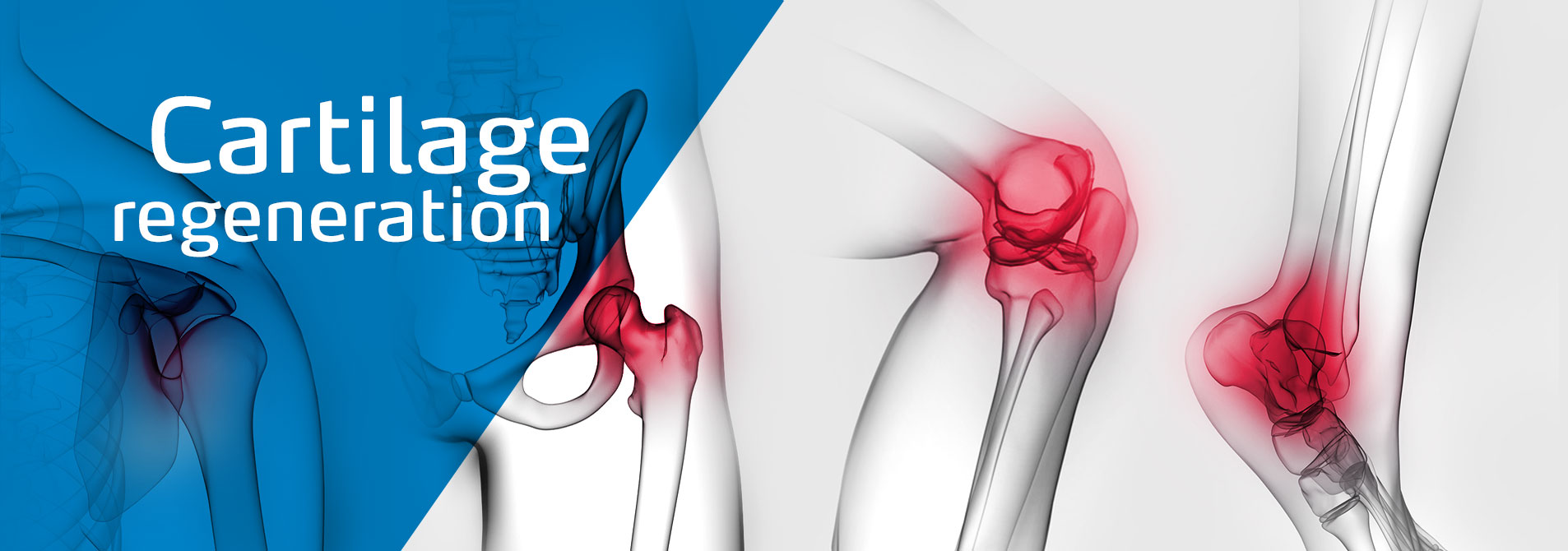
Cartilage damage (chondropathy) – when joints hurt
The bones that make up a joint are covered in smooth joint cartilage (articular cartilage). Together with the joint fluid (synovial fluid), the cartilage ensures that the joint can move with minimal friction and gently. In addition, the joint cartilage also has shock absorbing properties thanks to its compressive elasticity.
Every year, cartilage damage and bone cartilage damage afflict millions of people around the world. There are many reasons that can lead to it, but common causes include traumatic injuries or degenerative arthrosis due to the joint being overloaded or exposed to inappropriate stresses, as well as age-related wear and tear. Another cause is the bone disease osteochondrosis dissecans.
Depending on the severity, cartilage damage can be graded in several stages. A current and very widely used classification was developed by ICRS (International Cartilage Society).
ICRS classification of cartilage damage:
Since cartilage does not have its own blood supply, it is supplied via the synovial fluid and the subchondral bone. It follows from this that cartilage has almost no regenerative potential.
- Grade 0: Normal cartilage, no defects evident
- Grade 1: Superficial cartilage lesions: softening and/or superficial fissures/tears
- Grade 2: Lesions extending to < 50% of the cartilage depth
o Grade 3: Lesions extending to > 50% of the cartilage depth - Grade 4: Entire cartilage layer missing, subchondral bone is exposed
Which treatment technique? Determine the severity and cause of the defect
Cartilage damage should be treated in order to prevent it from getting worse. Today, a range of different therapy approaches are available. Which one is ultimately the best one depends on the severity and causes of the cartilage lesions.
“When it comes to cartilage damage, use regenerative therapies.”
Dr. Olaf. Th. Beck
Specialist in regenerative joint orthopaedics
Conservative treatment forms
With a conservative treatment it is possible to not only heal cartilage damage, but also to improve symptoms and slow down cartilage wear. The following conservative therapies are possible:
- Drug therapies (e.g. pain relieving drugs, hyaluronic acid)
- Physiotherapy
- Insoles and special footwear
- Aids such as bandages to relieve pressure and stress on the joint
Operative treatment methods
If surgical treatment is indicated, the choice of method depends on the size and depth of the defect. In addition, factors such as the intactness of the exposed bone, the quality of the cartilage on the opposite side to the defect, and the age of the cartilage defect all play a key role. The following surgical treatment techniques are possible:
Smoothing of the cartilage (“shaving”)
In this technique, cartilage components that have become detached or are in the process of detaching are surgically removed. The goal is to smooth off the cartilage surface so that inflammation processes in the joint can be reduced. This method can be used to alleviate the symptoms associated with certain cartilage lesions. However, the cartilage is not regenerated.
Bone marrow stimulation methods: microfracturing, nanofracturing, Pridie drilling
With bone marrow stimulation, the subchondral bone underneath the cartilage damage is opened with an awl, chisel or drill. As a result of these micro-injuries, blood from the bone marrow enters the cartilage defect. The pluripotent stem cells contained in this blood can form so-called repair cartilage (fibrous cartilage) in the damaged area. To support the process, an absorbable scaffold structure can be implanted in the cartilage defect, which serves as a matrix for the cells. In terms of biomechanical properties, repair cartilage is quite different to hyaline cartilage – it is softer and significantly less shock-resistant.
Autologous osteochondral transplantation (OCT)
Autologous OCT (autologous osteochondral transplantation) is a cartilage/bone transplant. Here, cylindrical plugs are removed from the bone cartilage from unloaded points on the same joint and grafted at the point of the cartilage defect. If multiple such cylindrical plugs are used then this is referred to as mosaicplasty. This method is limited, as only a few of these cylindrical plugs can be taken from healthy tissue. In addition, a defect is produced at the removal site, which can cause pain in some cases, even if it is outside of the loaded area. The gaps remaining between the plugs are filled with lower-quality scar tissue. Cartilage/bone plugs from the same joint are also referred to as osteochondral autografts. In contrast to this there are also osteochondral allografts, which are transplanted from multiple organ donors or deceased donors.
Autologous chondrocyte transplantation (ACT) / matrix-assisted autologous chondrocyte transplantation (MACT)
Two surgical procedures are required for cartilage cell transplantation. As the first step, cartilage biopsies are taken via arthroscopy from an unloaded area of the healthy joint cartilage. In the laboratory, cartilage cells are extracted from this tissue and multiplied. Afterwards the cultivated cartilage cells are inserted into the damaged cartilage in a second procedure. This technique is referred to as autologous chondrocyte transplantation (ACT). With matrix-assisted autologous chondrocyte transplantation (MACT), the isolated cartilage cells are implanted together with a three-dimensional scaffold matrix to promote cartilage regeneration.
Acellular matrix implantation / matrix-induced chondrogenesis (MIC)
As well as cartilage cell transplantation, there is also the gentler option of using a high-quality acellular biological matrix as an implant in the cartilage defect. The goal here is that endogenous cartilage cells and stem cells from the surroundings settle on the implant and are encouraged to form hyaline-like cartilage. At the same time, the exposed subchondral bone is protected with a protective matrix layer. In contrast to cartilage cell transplantation, this technique requires only one surgical procedure and involves no damage to cartilage through extraction of biopsies and no bone injuries due to microfracturing.
Artificial joint replacement
If the cartilage damage has progressed to a point where none of the techniques described above can be used, the last available therapeutic option for consideration is the implantation of an artificial prosthesis made of metal and plastic. However, the durability of artificial joints is limited. In addition, sufficient bone mass must be available to anchor the prosthetic. It must also be considered that prostheses cannot simply be replaced, as bone loss occurs after the first implantation. For this reason, this method should only be employed on older patients.
Cartilage damage
If cartilage damage goes untreated, this can often lead to premature arthrosis. If, for example, a piece of cartilage becomes completely detached, this can lead to blockage of the joint with acute strong pain and restricted mobility.
Local cartilage damage without an identifiable cause can occur particularly on the upper ankle joint, the knee and the elbow joint.
The year-on-year rise in joint disorders can be attributed to the following factors:
- Increase in life expectancy
- Increase in mass sports across all age groups
- Increase in higher-risk sports
- Increased mobility into old age
- Increase in body weight across the population
- Decrease in implantation of artificial joints on patients below the age of 60
Productinformation download:
ChondroFiller® liquid
product information leaflet (PDF)
ChondroFiller® liquid
patient information leaflet (PDF)
Facts about cartilage damage
The proportion of people who suffer from joint pain noticeably rises with increasing age. The joints most affected by this are the knee, shoulder and hip joints3.
Across Germany, around 5 million people suffer from complaints relating to the musculoskeletal system that require treatment. In 2018, around 230,000 of these people received inpatient surgical treatment (open surgery or arthroscopic surgery on joint cartilage and the menisci)4 .
Sources
1 Mithoefer K et al. Clinical efficacy of the microfracture technique for articular cartilage repair in the knee: an evidence-based systematic analysis. Am J Sports Med. 2009 Oct;37(10):2053-63.
2 Kreuz PC et al. Is microfracture of chondral defects in the knee associated with different results in patients aged 40 years or younger? Arthroscopy. 2006 Nov;22(11):1180-6.
3 Fuchs J, Prütz F (2017). Prävalenz von Gelenkschmerzen in Deutschland (Prevalence of joint pain in Germany). Journal of Health Monitoring 2(3): 66–71. DOI 10.17886/; RKI-GBE-2017-056.
4 DRG-Statistik 2018. Available online under: https://www.destatis.de/DE/Themen/Gesellschaft-Umwelt/Gesundheit/Krankenhaeuser/Publikationen/Downloads-Krankenhaeuser/operationen-prozeduren-5231401187014.pdf?__blob=publicationFile (17.03.2021).


